
Jaw Cysts and Benign Oral Lesions: How They’re Diagnosed and Treated
Most jaw cysts and benign oral lesions are discovered incidentally on X-rays, cause no immediate symptoms, and respond well to treatment — but the specific diagnosis determines everything about how they are managed.
Key Takeaways
- Jaw cysts and benign lesions range widely — from simple retention cysts to odontogenic keratocysts that require aggressive management.
- Diagnosis typically requires imaging (CT or panoramic X-ray) and often a biopsy.
- Treatment varies from enucleation (removal) to marsupializaton (decompression) to resection, depending on size, type, and location.
- Recurrence risk after treatment depends heavily on the specific lesion type.
What Jaw Cysts and Benign Lesions Actually Are
A cyst is a pathological cavity lined by epithelium, usually filled with fluid or semi-solid material. Benign oral lesions is a broader term encompassing any non-cancerous growth in the oral cavity, jaw, or surrounding tissue. The distinction matters clinically because they have different behaviors, recurrence rates, and treatment requirements.
Common Types of Jaw Cysts
- Dentigerous (follicular) cyst: Forms around an unerupted tooth’s crown; the most common odontogenic cyst.
- Radicular (periapical) cyst: Develops at the root apex of a non-vital tooth; almost always linked to pulp infection or necrosis.
- Odontogenic keratocyst (OKC): Aggressive cyst with high recurrence rates (up to 60% in some studies); associated with basal cell nevus syndrome in some patients.
- Lateral periodontal cyst: Found alongside root surfaces; typically discovered incidentally on X-rays.
Common Benign Oral Lesions
- Fibroma: The most common oral soft tissue tumor; typically caused by localized irritation or trauma.
- Torus (palatal or mandibular): Bony outgrowths in the palate or jaw floor; rarely require treatment unless interfering with function or dentures.
- Ameloblastoma: A benign but locally aggressive odontogenic tumor that can expand significantly and has high recurrence without adequate margins.
- Giant cell granuloma: Central or peripheral; central lesions are intrabony and require surgical intervention.
Many jaw cysts are secondary to untreated dental infections. Our article on why antibiotics cannot cure a tooth infection explains how unresolved pulp infections can lead to cyst formation over time.
Questions About Jaw Cysts Benign Oral Lesions
For Jaw Cysts Benign Oral Lesions, this part of Questions About Jaw Cysts Benign Oral Lesions focuses on the symptom history, previous treatment, current medicines, and the result the patient values most before treatment is scheduled, with the same details verified before treatment is scheduled. The clinician should also explain which examination finding supports each recommendation and what information could change it for Jaw Cysts Benign Oral Lesions when a second opinion is considered, using the examination and health history rather than a general assumption.
Clinical Checks for Jaw Cysts Benign Oral Lesions
For Jaw Cysts Benign Oral Lesions, this part of Clinical Checks for Jaw Cysts Benign Oral Lesions focuses on confirmed findings, measurements, imaging, and health-history factors when maintenance is discussed, with the same details verified before treatment is scheduled. The clinician should also explain what remains uncertain and whether another test or specialist opinion would alter the plan for Jaw Cysts Benign Oral Lesions during the initial review, using the examination and health history rather than a general assumption. Recording that explanation gives the patient a clearer next step for Jaw Cysts Benign Oral Lesions when maintenance is discussed, especially when follow-up duties are reviewed before treatment is scheduled.
Clinical Checks for Jaw Cysts Benign Oral Lesions During Planning
For Jaw Cysts Benign Oral Lesions, this part of Clinical Checks for Jaw Cysts Benign Oral Lesions During Planning focuses on confirmed findings, measurements, imaging, and health-history factors while comfort and function are assessed, with the same details verified before treatment is scheduled. The clinician should also explain what remains uncertain and whether another test or specialist opinion would alter the plan for Jaw Cysts Benign Oral Lesions while recovery expectations are clarified, using the examination and health history rather than a general assumption.
Clinical Checks for Jaw Cysts Benign Oral Lesions At Follow-Up
For Jaw Cysts Benign Oral Lesions, this part of Clinical Checks for Jaw Cysts Benign Oral Lesions At Follow-Up focuses on confirmed findings, measurements, imaging, and health-history factors before consent is finalized, with the same details verified before treatment is scheduled. The clinician should also explain what remains uncertain and whether another test or specialist opinion would alter the plan for Jaw Cysts Benign Oral Lesions before treatment is scheduled, using the examination and health history rather than a general assumption. Recording that explanation gives the patient a clearer next step for Jaw Cysts Benign Oral Lesions before consent is finalized, especially when follow-up duties are reviewed before treatment is scheduled.
Clinical Checks for Jaw Cysts Benign Oral Lesions For Long-Term Care
For Jaw Cysts Benign Oral Lesions, this part of Clinical Checks for Jaw Cysts Benign Oral Lesions For Long-Term focuses on confirmed findings, measurements, imaging, and health-history factors when the follow-up plan is written, with the same details verified before treatment is scheduled. The clinician should also explain what remains uncertain and whether another test or specialist opinion would alter the plan for Jaw Cysts Benign Oral Lesions as the long-term result is reviewed, using the examination and health history rather than a general assumption.
Clinical Checks for Jaw Cysts Benign Oral Lesions Before Treatment
Planning Jaw Cysts and Benign Oral Lesions: How They’re Diagnosed and Treated also means discussing effects on work, school, travel, eating, driving, exercise, and caregiving responsibilities. Before scheduling Jaw Cysts and Benign Oral Lesions: How They’re Diagnosed and Treated, ask how delays, missed visits, or slower healing could change comfort, cost, and the final result before consent is finalized.
The Diagnostic Process
Imaging
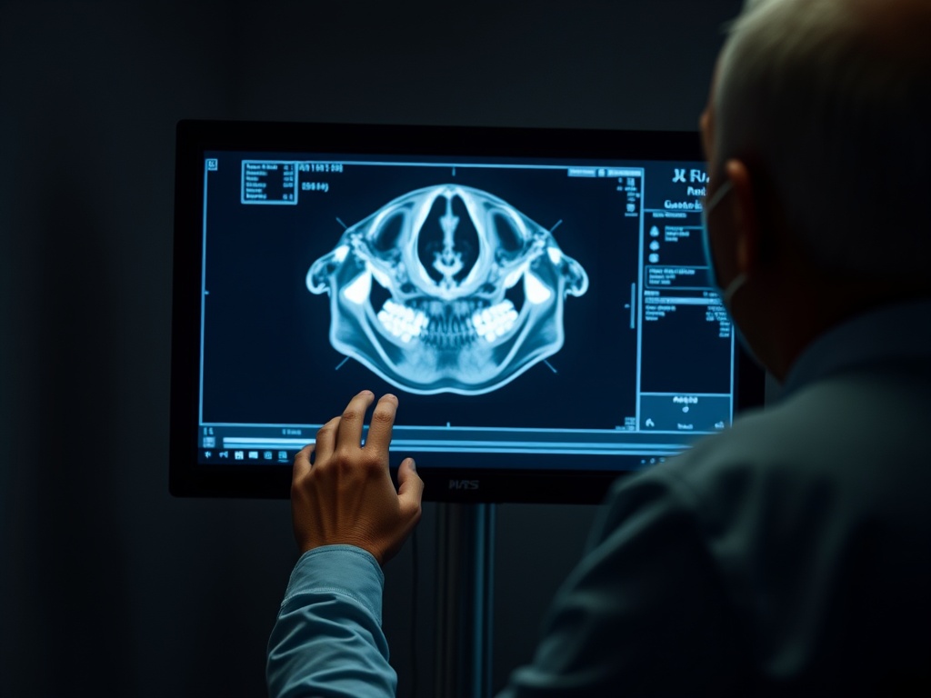
Panoramic X-rays provide the initial map. Cone-beam computed tomography (CBCT) adds three-dimensional detail about lesion extent, relationship to adjacent teeth and nerves, and cortical plate involvement. According to the American Academy of Oral and Maxillofacial Radiology, CBCT is particularly valuable for large or complex jaw lesions before surgical planning.
Biopsy and Histopathology
Most jaw lesions require tissue biopsy before definitive treatment. This may be an incisional biopsy (a sample taken from a larger lesion) or an excisional biopsy (the entire lesion removed and submitted). Histopathology determines the exact diagnosis and drives the treatment decision.
Surgical Decision Points
Treatment choice depends on lesion type, size, anatomic location, and the patient’s overall health. The primary surgical options are:
| Procedure | What It Involves | Best Suited For | Recurrence Risk |
|---|---|---|---|
| Enucleation | Complete removal of cyst lining | Small to moderate cysts with clear margins | Low for most cysts; higher for OKC |
| Enucleation + Carnoy’s solution | Chemical fixation of remaining lining | OKC specifically | Reduced vs enucleation alone |
| Marsupialization | Surgically opening cyst and allowing it to drain and shrink over time | Very large cysts near vital structures | Higher; often followed by secondary enucleation |
| Peripheral ostectomy | Removal of a rim of surrounding bone | OKC, selected aggressive cysts | Reduces recurrence compared to enucleation alone |
| Resection | Segmental jaw removal with reconstruction | Aggressive tumors (e.g., ameloblastoma) | Lowest for aggressive lesions |
Specialist Referral: When and Why
General dentists often identify jaw cysts but typically refer to oral and maxillofacial surgeons for:
- Any lesion larger than 2 cm or crossing anatomic boundaries
- Lesions with features suggesting aggressive behavior on imaging
- Recurrent cysts after prior treatment
- Cases where the lesion is near the inferior alveolar nerve, maxillary sinus, or other critical structures
If dental anxiety is a barrier to pursuing evaluation or surgery, our guide on trauma-informed dental care discusses approaches that oral surgeons and dental teams can use for patients with difficult past experiences.
Long-Term Monitoring After Treatment
Recurrence surveillance is essential, particularly for OKC, ameloblastoma, and giant cell lesions. Most oral surgeons schedule:
- Clinical and radiographic review at 6 months post-surgery
- Annual panoramic or CBCT imaging for 3–5 years
- Longer-term follow-up for aggressive subtypes
Lesions discovered and treated early typically have straightforward recoveries. The concern is the lesion found late — after significant bone expansion, nerve displacement, or adjacent tooth involvement. For any unusual findings on routine dental X-rays, a prompt specialist referral is the most protective next step.
Questions about whether a consultation can happen virtually before committing to in-person specialist care are addressed in our overview of virtual vs in-person dental consultations.
[TABLE]